What is this a photo of?

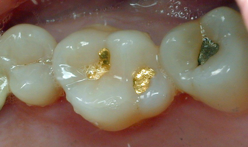
Something I see far too often lately! This is a broken ceramic crown on a patient’s lower left second molar. It fractured in two, and the half on the tongue side of the molar is still hanging on. The patient thinks she had the crown placed about eight years ago.
Why did this happen?
The human jaw is a lever system configured like a nutcracker. Your muscles can generate about four times the force in the second molar area than can be generated on your incisor teeth in front. Some patients grind and clench their teeth. It is no coincidence that most fractures of teeth occur on second molars.
The emphasis on esthetics nowadays is so powerful that some patients think I am crazy when I tell them second molars are not the place for ceramic crowns. They either break like this one did, or repeatedly come uncemented. If you have to drill a hole through a ceramic crown to do a root canal, good luck to you. It is a tough job. Can you imagine drilling a hole through a porcelain dinner plate?
Computer-milled zirconia crowns were invented almost twenty years ago. Like most new products, their advantages were exaggerated. They were tougher than traditional porcelain crowns with a metal substructure, it is true. But they were NOT unbreakable, as was claimed. It was advertised that they could be made as thin as half a millimeter, like metal crowns, and still resist fracture. That turned out to be a lie.
Two other disadvantages of milled zirconia crowns: they are so inert that cement often does not bond well to them, and they are so hard that they often wear the natural tooth above or below them.
When I tell patients these facts, the response is often, “Why didn’t my former dentist give me other options?”
The reason is that computer-milled zirconia crowns are currently the cheapest ones available from dental labs. Low cost drives their popularity with dentists and patients alike. But how much does a patient save if a ceramic crown breaks in a few years and has to be replaced at additional expense?

What are the alternatives to ceramic crowns?
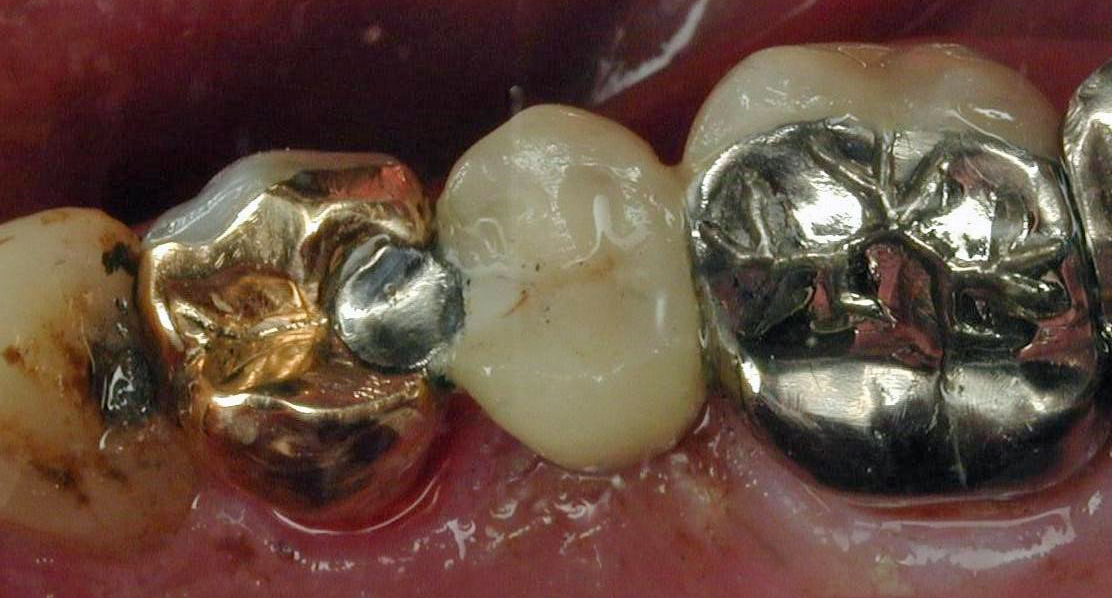
They may not be white, but I have never in my career seen a cast metal crown break in two. For nearly a century, metal crowns, particularly ones made of gold alloys, were seen as the standard of care for posterior teeth. They are the crowns dentists themselves most often have in their own mouths!
Nowadays gold is considerably more expensive, but we often make crowns out of more economical alloys like silver-palladium and chrome-cobalt alloys, and they are just as durable.
In the old days when gold crowns were the norm, teeth were cut down far more conservatively, and as a result, fewer teeth needed root canals. Gold crowns occasionally wore through on the chewing surface, but that could be repaired. A fractured ceramic crown cannot be salvaged!
As I tell patients every day, durability needs to be factored into patient choices in dental treatment. Especially in high force areas like the second molar region, it is hard to beat cast metal crowns. Sometimes the tried-and-true methods of doing things are still the best way!
Kim Henry, DMD
December 5, 2023


 rents of a 13-year-old child came in to have me review the appropriateness of their son’s orthodontic treatment. The father had wanted to bring their son to me for treatment originally, but the mother saw an advertisement for a chain of orthodontic clinics, promising substantial savings. The mother’s wish prevailed, and the child was 1 year into orthodontic treatment at this clinic with little progress.